Gel Imaging System
GBOX EF3
Description
The G:BOX F3 has a high resolution 3.8m pixel camera which is capable of giving outstanding images with incredible spatial resolution. The system is suitable for a wide range of standard fluorescence applications such as DNA imaging or protein gels.
Features
| |
 |
Compact darkroom with sliding door - has a fully light tight, robust darkroom suitable for advanced chemiluminescence, fluorescence and visible light applications. |
| |
|
No compromise on resolution - Ultra quiet cameras ranging from 1.4m up to 8.3m pixels. Some cameras have varying degrees of cooling. |
| |
|
A 7 position computer controlled filter wheel is standard |
| |
|
All lenses are computer controlled and are motor driven |
Advantages
| |
|
DNA detection with Ethidium Bromide or 'Safe' dyes - Using a 302nm UV transilluminator and Orange emission filter, images of ethidium bromide stained DNA gels can be captured in a fraction of a second. |
| |
|
Protein analysis - Protein gels stained with either Coomassie blue stain or silver stain can easily be captured with a G:BOX using a visible light converter screen. |
| |
|
Chemiluminescence with or without colourimetric markers* - Chemiluminescence with a G:BOX is very easy. Simply place the blot inside the system and GeneSys will assess the output |
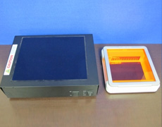
U Genius 3
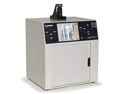
Designed to make your gel imaging simple, quick and easy. No set up, no external computer, just a complete imaging system for all your 1D needs. U:Genius3 – from switch-on to perfect image at a touch. It is the perfect choice for low budget gel documentation and analysis fluorescence applications.
Features
| |
|
Compact darkroom with sliding door - 46.5(w) x 51.0(h) x 39.0(d) cms |
| |
|
Can use a range of transilluminators (20 x 20 cm) |
| |
|
No compromise on resolution - 3 million pixels |
| |
|
Easy to access filter drawer accepts interchangeable filters |
| |
|
Integral computer |
| |
|
Colour touch screen |
Advantages
| |
|
High specification - The system features a high resolution camera with 3 million pixels. It has a manually operated zoom lens and a filter drawer system which accepts interchangeable filters for a wide range of applications. |
| |
|
Built in processor - An integral computer running embedded Windows controls the system using the larger touch panel display. |
| |
|
U:Genius3 software - The intuitive "button controlled" software is simple to use. Camera and exposure control is via the touch screen. It is also possible to make image enhancements and annotations from the touch screen. |
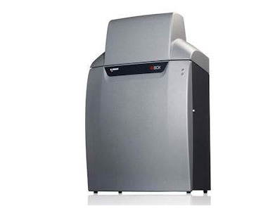
GBOX ICHEMI

Description
G:BOX Chemi systems provide the user with an extended range of applications covering fluorescence and chemiluminescence. The G:BOX Chemi series features cameras with resolutions from 1.4m pixels up to 8.3m pixels. All have cooling which reduces the small amount of electronically generated noise to produce outstanding images with minimal background interference.
Features
| |
|
Compact darkroom with sliding door - has a fully light tight, robust darkroom suitable for advanced chemiluminescence, fluorescence and visible light applications. |
| |
|
No compromise on resolution - Ultra quiet cameras ranging from 1.4m up to 8.3m pixels. Some cameras have varying degrees of cooling. |
| |
|
A 7 position computer controlled filter wheel is standard |
| |
|
All lenses are computer controlled and are motor driven |
Advantages
| |
|
DNA detection with Ethidium Bromide or 'Safe' dyes - Using a 302nm UV transilluminator and Orange emission filter, imagesof ethidium bromide stained DNA gels can be captured in a fraction of a second. |
| |
|
Protein analysis - Protein gels stained with either Coomassie blue stain or silver stain can easily be captured with a G:BOX using a visible light converter screen. |
| |
|
Chemiluminescence with or without colourimetric markers* - Chemiluminescence with a G:BOX is very easy. Simply place the blot inside the system and GeneSys will assess the output |
Benefit
| |
|
Performance - With the new GeneSys control software the user simply has to tell the G:BOX Chemi the sample type and dye and...(Missing Text) |
Proteomics
Syngene, with its imaging expertise, has developed high specification equipment and software for challenging 2D electrophoresis gel imaging, to bring you the Dyversity systems, Proteomesan and Dymension software.
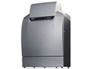
2D IMAGING SYSTEM (DYVERSITY)
| |
|
Dyversity is designed for the rapid generation of 2D protein gel images and features a high resolution, 16-bit CCD camera inside a computer-controlled darkroom. This camera offers unrivalled separation of the highest density of protein spot images and can capture faint spots guaranteeing imaging accuracy which is comparable to many expensive laser based scanning systems. |
| |
|
Dyversity system has a motor driven sample stage, motor driven lens and can be fitted with a range of filters, UV and visible lighting options to allow imaging of all commercial 2D protein stains including Coomassie Blue, silver stain, SYPRO® Ruby, Pro-Q® Diamond, Deep Purple™. With an optional lighting module it is also possible to image gels stained with Cy2, Cy3 and Cy5. |
|
 |
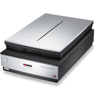
PROTEOMESCAN
| |
|
Proteomescan system can generate high quality 2D gel images in seconds which can be automatically transferred into Dymension, Syngene's revolutionary 2D image analysis software. |
| |
|
Proteomescan users can switch between transmittance and reflectance modes and can select required resolution. The system can generate images of up to 12,800 x 12,800-dpi resolution and has variable colour scanning, which makes it easy to scan gels stained with coloured dyes such as Coomassie Blue and silver stain and ensures detection of the smallest protein features. |
|
 |
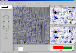
DYMENSION
| |
|
Dymension is revolutionary software that can analyse a typical 2D gel image rapidly. It features novel algorithms for background subtraction, noise filtering, precise alignment, spot detection, rapid matching and reduced image editing time. Using its powerful spot detection algorithm, Dymension instantly locates and analyses protein spots. With Dymension, the entire analysis process from background correction to spot matching results and reporting takes minutes, making this the fastest 2D gel analysis package currently available. |
|
 |
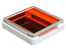
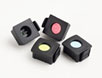
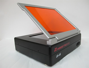
UltraSlim LED Illuminator
Product Summary
Designed for viewing and cutting stained gels on laboratory bench, or within gel documentation systems. The dimensions is only 210(W) x 210(D) x 30(H)mm, which is space saving and the THINNEST in the world.
| |
|
Highly sensitive as it has been optimized for use with most nucleic acid and protein stains, sensitivity up to 0.5ng |
| |
|
User friendly for gel viewing and cutting |
| |
|
Simple as no amber glasses nor protective outfit needed |
| |
|
Safe as blue LEDs does not cause damage to eyes and skin and NO DNA damage due to UV exposure |
| |
|
High uniformity with less than 10% coefficient variance |
Features
| |
|
Compact (30mm in height) |
| |
|
THINNEST in the world |
| |
|
Enlarge laboratory space |
| |
|
Feather like weight for mobility |
| |
|
Robust and stylish design |
More Info:
http://www.maestrogen.com/product_detail.asp?Pid=53
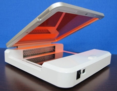
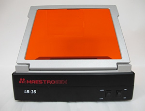
UltraBright LED Transilluminator, 470NM
Catalog Number: LB-16 |
|
 |
Product Summary
Designed for viewing and cutting stained gels on laboratory bench, or within gel documentation systems. The dimensions is only 210(W) x 210(D) x 30(H)mm, which is space saving and the THINNEST in the world.
| |
|
Safer alternatives to replace carcinogenic UV transilluminator |
| |
|
Ease of use for gel cutting and gel viewing |
| |
|
Flexible-Adjustable intensity from 100% down to 50% light intensity |
| |
|
Strong blue LED excitation |
| |
|
Simple-amber glasses and protective glove eliminated |
| |
|
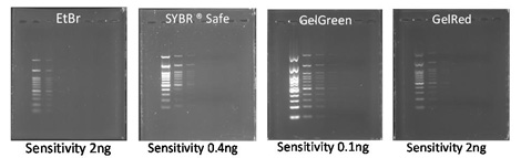
Compatible with a wide range of nucleic acid stains and protein stains: EtBr, SYBR® Safe, SYBR Gold, SYBR® Green I & II, SYPRO® Ruby, SYPRO® Orange, Coomassie Fluor™ Orange stains, GelGreen, GelRed and Lumitein™ Protein Gel Stain |
Features
| |
|
Amber filter integrated |
| |
|
Large viewing surface—160D x 200W (mm) |
| |
|
LED life up to 50, 000 hours |
| |
|
High intense uniform blue LED's light |
More Info:
http://www.maestrogen.com/product_detail.asp?Pid=42
|